◆中国科技核心期刊
◆中国应用型核心期刊
◆中国医药卫生核心期刊
◆中国高校优秀科技期刊
◆中国应用型核心期刊
◆中国医药卫生核心期刊
◆中国高校优秀科技期刊
◆美国《化学文摘(CA)》收录
◆美国《剑桥科学文摘(CSA)》收录
◆波兰《哥白尼索引(IC)》收录
◆日本科学技术振兴机构数据库(JST)收录
◆美国《剑桥科学文摘(CSA)》收录
◆波兰《哥白尼索引(IC)》收录
◆日本科学技术振兴机构数据库(JST)收录
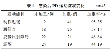
Objective : To investigate the changes of peripheral blood inflammatory markers in patients with Parkinson’s disease (PD) after severe acute respiratory syndrome coronavirus 2 ( SARS-CoV-2) infection , and to analyze their correlation with the exacerbation of different motor symptoms. Methods: A total of 43 PD patients with SARS-CoV-2 infection admitted to Affiliated Hospital of the Institute of Neurology, Anhui University of Chinese Medicine from November 2022 to May 2023 were enrolled. The Unified Parkinson’s Disease Rating Scale- Ⅲ (UPDRS- Ⅲ) was used to assess the aggravation of motor symptoms before SARS-CoV-2 infection (within one month before admission without infection) and after infection (within 7 days after diagnosis) , and the results were compared. Peripheral blood inflammatory indicators of the patients, including neutrophil count (NEUT) , lymphocyte count (LYM) , neutrophil-to- lymphocyte ratio (NLR) , C-reactive protein (CRP) , high-sensitivity C-reactive protein (hs-CRP) , and IL-6, were collected. Correlation analysis was conducted to explore the relationship between these indicators and the aggravated motor symptoms, and Logistic regression analysis was performed to screen the independent risk factors for the aggravation of motor symptoms in PD patients after SARS-CoV-2 infection. Results: Compared with the UPDRS- Ⅲ score before infection [ 15. 00 ( 11. 00, 22. 00 ) points] , the UPDRS- Ⅲ score after infection [ (27. 74±8. 08) points] was significantly increased (Z = -5. 716, P < 0. 05 ). Correlation analysis showed that NEUT, NLR, CRP , and IL-6 were positively correlated with symptom aggravation (r=0. 463, 0. 556, 0. 505, 0. 453, all P<0. 05). Logistic regression analysis revealed that NEUT, NLR, CRP , and IL-6 were independent risk factors for the aggravation of muscle rigidity(OR= 1. 289, 1. 340, 1. 025, 1. 061, all P<0. 05) ; NEUT, CRP , and IL-6 were associated with the aggravation of tremor (OR = 1. 204, 1. 027, 1. 051, all P<0. 05) ; and CRP was the risk factor for postural reflex disorder ( OR = 1. 030, P = 0. 019 ). Conclusion: In PD patients co-infected with SARS-CoV-2, motor symptoms significantly worsen, accompanied by marked elevations in NEUT, CRP , and IL-6 levels, which are closely associated with increased muscle rigidity and tremor severity.